Blinded with Science: DXA Scan
 Last week I made my way to the University of Washington General Clinical Research Center (GCRC) to get a DXA scan. This wasn't quite a research study, but was part of the training for a DXA technician.
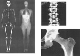
A DXA scan is essentially a full-body X-ray. In the case of a normal X-ray, one set of frequencies is used in order to view skeletal structures, but the DXA scan uses two sets, one stopped by bone, the other by soft tissue. By examining the differences between the amounts absorbed of each frequency, the DXA scan can give a precise measurement of bone mineral density. This scan is primarily used on post-menopausal women to help in diagnosis and tracking of osteoporosis. Young, healthy males aren't prone to issues with bone density loss, so it's a bit harder for technicians to get that experience, putting me in the "desired" demographic for someone learning to do the scan.
After walking the maze that is the UW Hospital, I eventually found the GCRC. They took my measurements (showing that I'm both heavier and shorter than I'd like), and then it was time for the scan. You know how the dentist places your head in order to get a good X-Ray? Imagine that with your entire body. Getting a good DXA scan is dependent on having the body aligned with defined reference points. Once aligned, all you have to do is stay still, feeling a bit like a piece of paper in a copier. There were three tests done, one full body, one for the lumbar spinal area, and the last for the hip. The latter two areas are areas where bone density loss shows up first.
So what did I get out of this experience other than a nice dose of radiation? Data. Pages and pages of data. While bone density measurement is the primary use, a DXA scan also gives other information regarding body compostion, namely body fat percentage. It even breaks that down further into different areas, so I now know that I have body fat symmetry (and where that tends to be held). Interesting stuff indeed. Knowing all of that has given me a motivation for another personal spanning_time event: finding a gym.

0 Comments:
Post a Comment
<< Home